
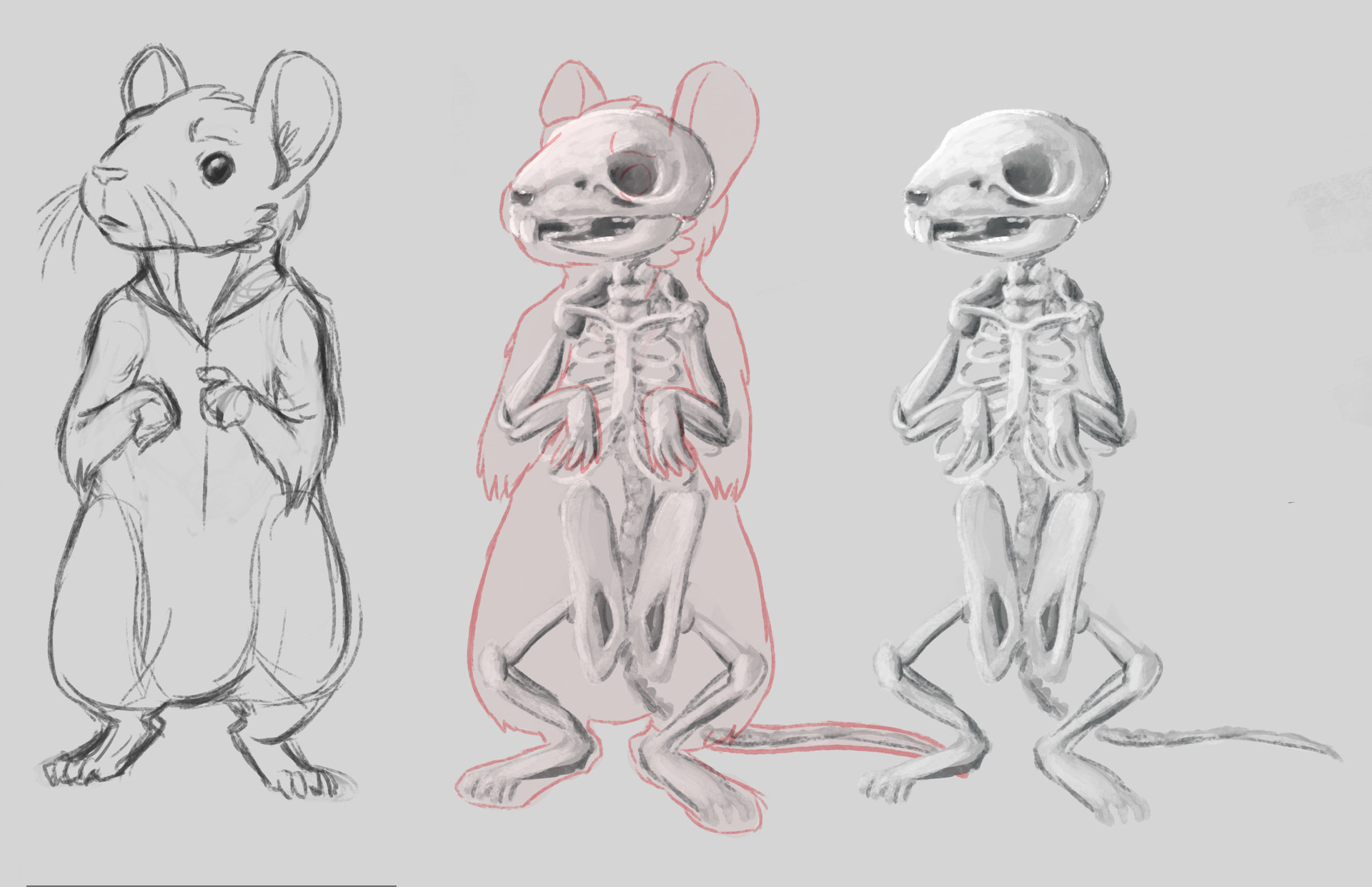
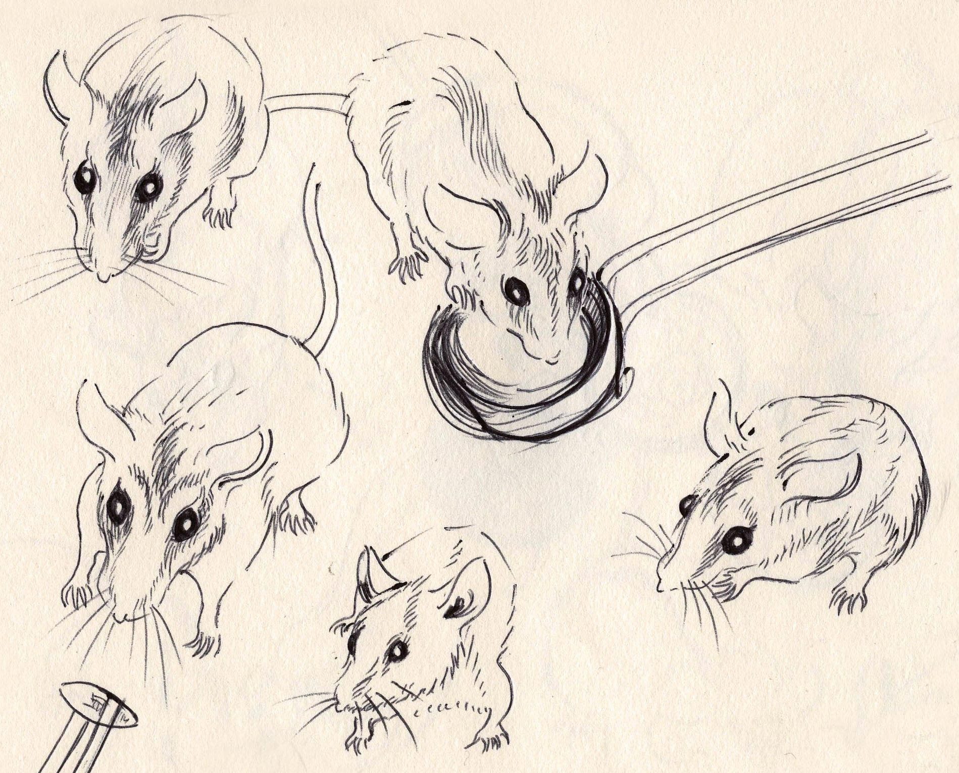
rowkey “ Some quick mouse form/anatomy practice gave ‘em BEES. Definitely more to come. 8
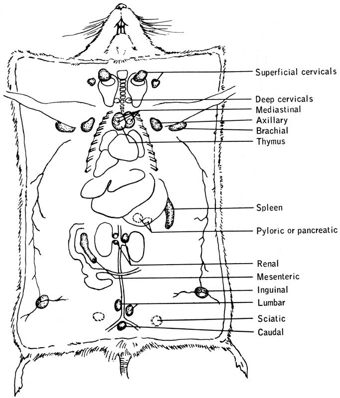
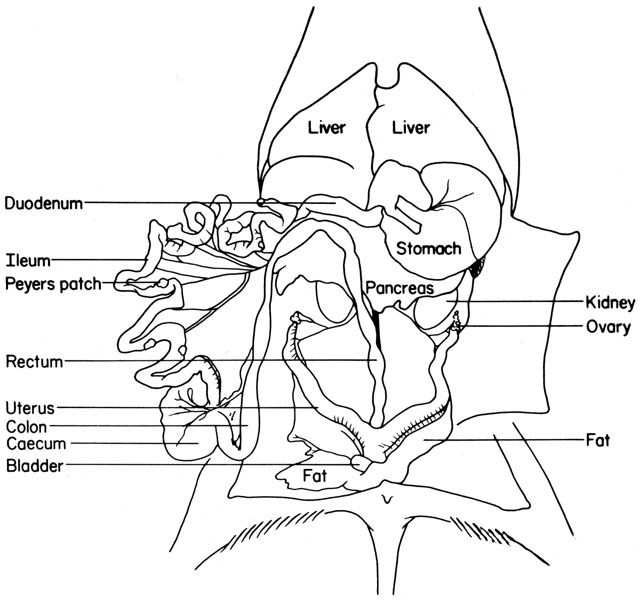
In general, images in these atlases are camera lucida-based line drawings rather than accurate three-dimensional images. Furthermore, so far a systematic two- and three-dimensional description of the internal anatomy of bones, as well as the three-dimensional relationship exhibited in joints, are not available.
Mouse Anatomy Anatomical Charts & Posters
Home : Mouse Anatomy Rat and Mouse Anatomy Rat and Mouse Anatomy Comparative Anatomy of the Mouse and Rat: a Color Atlas and Text provides detailed comparative anatomical information for those who work with mice and rats in animal research. Order your anatomy atlas from the AALAS Store!
Anatomy Of Mice
The mouse remains the key animal model for exploring human disease and, despite its small comparative size, the laboratory mouse is anatomically similar to humans, providing even unexpected anatomical analogies in structures with high interspecies variation such as the presence of the clavicle.
The Anatomy of the Laboratory Mouse
The Anatomy and Physiology of Laboratory Mouse Sarita Jena & Saurabh Chawla Chapter First Online: 24 July 2021 3660 Accesses Abstract Among the different types of vertebrate and invertebrate animals used in biomedical research, the laboratory mouse is the widely used vertebrate animal model.
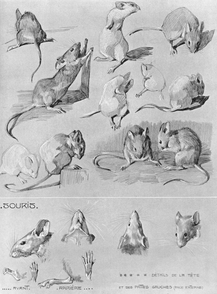
Mouse Drawing Reference and Sketches for Artists
illustration labeled labelled labels male mammal model organism mouse mus musculus nature no-one
Biology of the Laboratory Mouse Figure 1319
One chapter on mouse anatomy by Komárek V. Drawings taken from Popesko et al. (2002) A Practical Guide to the Histology of the Mouse 2014 Scudamore CL Willey Blackwell Histological text and atlas with the basis of mouse anatomy Morphological Mouse Phenotyping. Anatomy, Histology and Imaging 2016 2017 Ruberte J, Carretero A, Navarro M Editorial.

The Anatomy of the Laboratory Mouse
It guides through normal mouse anatomy and histology using direct comparison to the human. The side-by-side comparison of mouse and human tissues highlights the unique biology of the mouse, which has great impact on the validation of mouse models of human disease.. Source: Drawing by Dr. S. Chou, Charles River, with permission. C57BL/6 Mice.

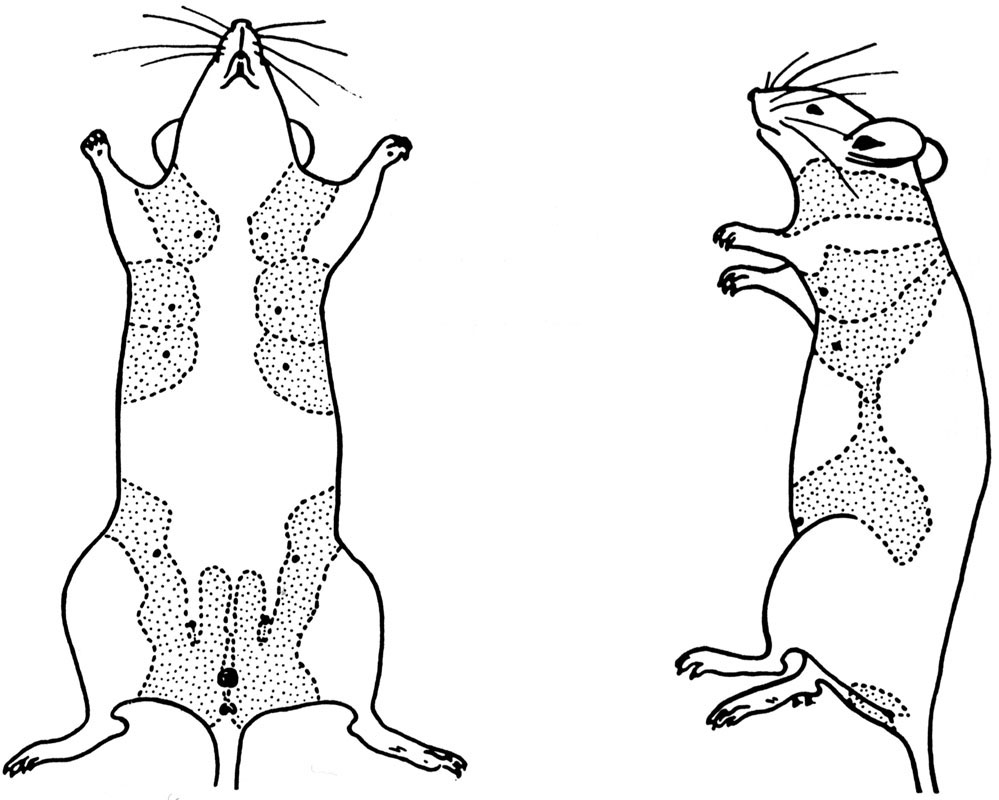
Male mouse anatomy Illustration by Laurie O'Keefe Medical Illustration & Animation
The floating ribs are not drawn. 31. Right scapula and clavicle. 32. Thoracic cage, formed by 13 thoracic vertebrae, 13 pairs of ribs and the sternum. 33. Lateral aspect of right humerus, radius and ulna. 34. Flexor surface of right humerus.
Mouse Drawing Reference and Sketches for Artists
3-D WIRE MODEL BASED ON MRI SECTIONS Quicktime Mouse Radiographic Atlas of Skeletal Anatomy The following link will take you to a series of radiographic images with color overlays and labels. To proceed click here. Comparative Anatomy Chart This table contains a comparison between mouse and human anatomy.

Rowkey — Some quick mouse form/anatomy practice gave ‘em... Pencil Drawings Of Animals, Animal
Mouse Anatomy C57BL/6 mouse embryo -- Anatomy Marked (click thumbnails to enlarge) FaceBase is the primary data resource for craniofacial researchers worldwide.

rat. mouse? Anatomy art, Human figure drawing, Anatomy drawing
The mouse remains the key animal model for exploring human disease and, despite its small comparative size, the laboratory mouse is anatomically similar to humans, providing even unexpected anatomical analogies in structures with high interspecies variation such as the presence of the clavicle.

Mouse anatomy, illustration Stock Image C047/1854 Science Photo Library
The Mouse Limb Anatomy Atlas is a free, web-based, standardised reference of limb muscle, tendon and skeletal structures at embryonic day 14.5. The Atlas features interactive and annotated 2D and 3D models of the forelimb and hindlimb, showing over 60 individually segmented structures. This is the first complete reference tool for studying the.
The Anatomy of the Laboratory Mouse
HOME vet-Anatomy Mouse - Whole body Labeled cross-sectional anatomy of the mouse on micro-CT Antoine MICHEAU, MD , Denis HOA, MD Authors affiliations Publication date: May 30, 2018 | Last update: Sep 23, 2022 https://doi.org/10.37019/vet-anatomy/564757 ISSN 2534-5087

Studies of See Through Mice Animal sketches, Animal drawings, Mouse illustration
Item Details: The Mouse Anatomy Poster drawings were done by Gheorghe M. Constantinescu, DVM, PhD, Drhc, a veterinary anatomist and medical illustrator at the University of Missouri-Columbia. 06-00018: This poster is a valuable reference tool for researchers and laboratory technicians, as well as an excellent addition to high school or college.
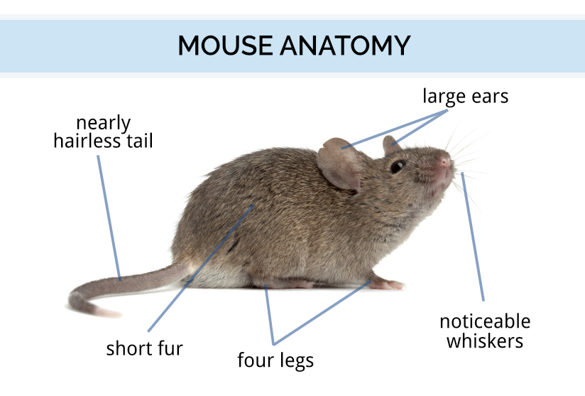
Mouse Identification & Anatomy How Long Mice Live
We originally obtained vector drawings of Nissl 2D section from Paxinos and Franklin's the Mouse Brain in Stereotaxic Coordinates, 3rd edition 6. We also used the 4th version to incorporate the.
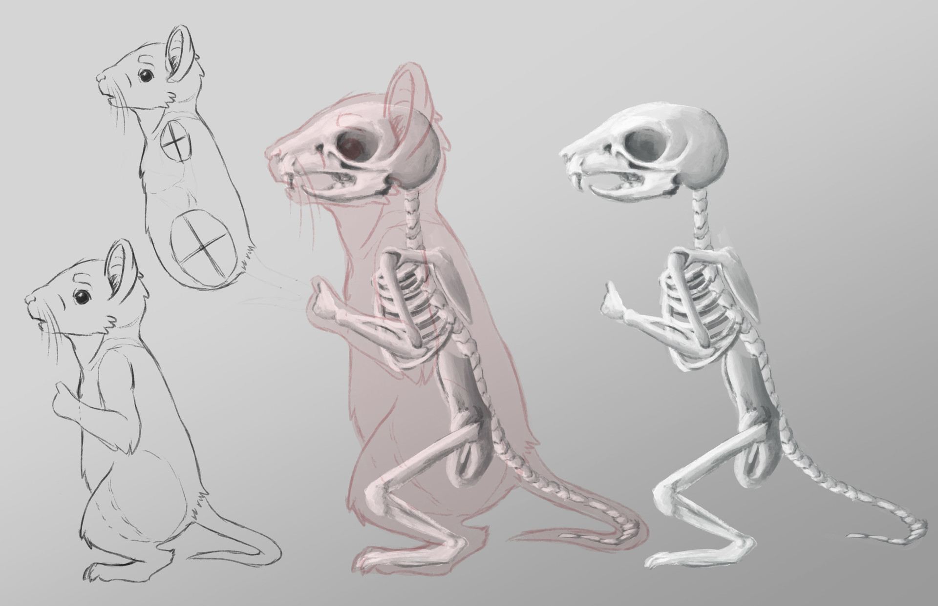
ArtStation Redwall Developing Mice
We analyzed the mouse whole-body model and described the moment-arms for different hindlimb and forelimb muscles, the moments applied by these muscles on the joints, and their involvement in limb movements at different limb/body configurations.